Tita Karlita Designed a 3D Bone Ultrasound Imaging System for Her Dissertation
TIMESINDONESIA, SURABAYA – Tita Karlita, a Doctoral student of Department of Electrical Engineering, Faculty of Electrical Engineering and Intelligent Informatics, designed a 3D Bone Ultrasound Imaging System to reconstruct bone outer counters for her dissertation.
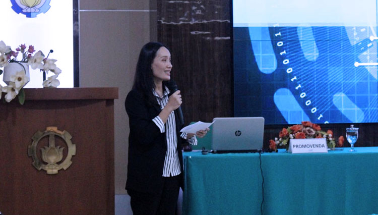
The dissertation was delivered at Institut Teknologi Sepuluh Novemeber (ITS) on her Open Session for Doctoral Program on Tuesday (2/25/2020).
Advertisement
On her dissertation, Tita explained how the 3D Ultrasound Imaging System could be a better choice to minimize the area of bone detection.
"So far Computerized Tomography (CT) is recognized as the best standard in bone imaging. However, the method that uses X-ray has high radiation, so the frequency of use is limited," Tita said on Friday (2/28/2020).
In addition she also explained that ultrasound is currently not recommended for bone imaging. However, ultrasound has the advantage that it does not emit radiation and its cheaper.
"NEURO is a combination of two methods, namely Regent Proposal Network (RPN) and Curve Approximation," she added.
Furthermore she also explained that on the implementation, 3D Ultrasound only perform scanning and reconstruction without segmentation, while NEURON tries to eliminate disorders such as muscles or tendons so that the purpose of bone contour reconstruction can be accomplished.
This 3D Bone Ultrasound Imaging System delivered Tita Karlita to achieve her doctoral degree at the Department of Electrical Engineering, Faculty of Electrical Engineering and Intelligent Informatics of Institut Teknologi Sepuluh November (ITS).
**) Ikuti berita terbaru TIMES Indonesia di Google News klik link ini dan jangan lupa di follow.
| Editor | : Khodijah Siti |
| Publisher | : Lucky Setyo Hendrawan |